Sunday, May 10, 2020

Electrophoresis

Take a rectangular tank and epoxy in electrodes at opposing ends, like this


Add an appropriate buffer, and then hook the electrodes up to + and - output from a regulated power supply, and you will get a voltage between the two wires that causes a current to flow.  Biological molecules carry electric charge, so they will move too, in an appropriate supporting medium.

Electrodes are made from thin platinum wire.  Since the molecules are negatively charged, they move toward the cathode (red).


Gels

Two materials have traditionally been used for gels:  polyacrylamide and agarose.  Acrylamide is on the left, and its polymerized form is on the right.

 
Acrylamide is cross-linked into a mesh by the inclusion of a small amount of bis-acrylamide (technically, N,N' methylene-bisacrylamide).


You mix a solution of acrylamide and bis-acrylamide plus the appropriate buffer, then the reaction is started by addition of a small amount of TEMED and ammonium persulfate (5-20% is a range for acrylamide).  The mixture is poured into a mold (glass plates separated by spacers, with something at the bottom to keep the liquid from running out.

Once it's set, the plug at the bottom is removed.  Electrical continuity is maintained by wedging a piece of sponge into the bottom.

A gel mold contains two glass plates, one of which is notched.  The "ears" of the notched plate have a tendency to get broken, so usually a set comes with an extra notched plate when you buy them.

The other material is agarose.  This is a purified form of the agar that is used for bacteriological plates (petris dishes).  Agar is a polysaccharide extracted from certain kinds of seaweed.  Agar has been used to solidify desserts for a long time, it was introduced to Koch's laboratory by the wife of one of his assistants, who knew about it.  He never publicly credited her with the idea.

[ It's amusing that the very first medium used for isolation of single colonies of bacteria was a potato, sliced through.  Appropriate for a German laboratory, I think. ]

Biochemically agarose is a repeating polymer of dimers of galactose plus a galactose derivative.

Agar and agarose have the property that when mixed with water, boiled and then cooled, the material sets into a gel (like jello, but generally stiffer) at around 45°C.  Once set, it can be heated a lot higher without losing its physical properties.  The stiffness depends on the concentration of agarose used.  0.8-1.5% would be usual

Agarose gel electrophoresis also requires an appropriate buffer (e.g. Tris-acetate).  This type of electrophoresis is extremely convenient.  The gel is non-toxic, easily prepared by boiling, and the gel can be poured flat (see the picture above).  You can't do this with polyacrylamide because oxygen inhibits the polymerization reaction.

Samples are loaded into wells under the surface of the buffer.  The aqueous samples are made dense by addition of glycerol to 10% or so.  Dyes (bromophenol blue and sometimes xylene cyanol) are also added into the samples, they move at characteristic rates and allow you to visualize the progress of the separation.

Separation in electrophoresis

For DNA at neutral pH, charge is carried by the phosphate groups, which contribute 1 or 2 negative charges (the average depends on the exact pH).  This means that the charge to mass ratio is constant for DNA or RNA of different lengths.

The reason that DNA or RNA molecules of different sizes separate is the existence of a retarding force that is greater for larger molecules.  Or maybe it's better to turn that around:  we observe that the log of the distance traveled is inversely proportional to the length of the molecule, and infer the existence of a force that depends on length.

Samples for protein gels are typically prepared by boiling a protein mixture in the presence of a detergent SDS (sodium dodecyl sulfate).  The hydrophobic part coats the protein and destroys its secondary structure.  The evenly spaced sulfate groups impart negative charge.  As with DNA,  the charge to mass ratio is constant for polypeptides of different lengths.  Separation occurs by means of the size-dependence of the retarding force.

Visualization

The classic method for visualizing protein gels is to stain with a blue dye (Coomassie brilliant blue).  In this picture we can see a protein gel drying after the electrophoresis has been run.  The blue spots are proteins.

DNA gels were often stained with a fluorescent dye such as ethidium bromide.

Ethidium is moderately mutagenic, so substitutes have been developed.

Alternatively if the material is radioactive, you just expose the dried (or even wet) gel to X-ray film.  These days, they have fancy apparatus that records the emitted beta particles without the use of film.  I remember the revolution caused by the introduction of automatic film processors.

Laemmli

To get the best resolution, you want the bands of protein or nucleic acid to be as thin as possible.  Here is a gel with very nice resolution:


The thickness of the bands depends on how much of each protein is present in the sample.

To get a pretty gel (one with nice thin bands), for DNA or RNA the important thing is to have as little sample as possible and to run a thin gel (like 0.4 mm).

For protein gels, there is a trick, invented by Laemmli.  There is a combination of two gels, one on top called the stacking gel, and a larger one below called the running gel  The system has 3 different buffers.  The upper and lower tank buffers contain glycine as the mobile anion and are at pH 8.8.  The gels are

Stacking gel:  3% acrylamide, pH 6.8
Running gel:  5% - 20% acrylamide, pH 8.8

This system compresses a sample which might be almost a centimeter from top to bottom when first loaded, into a set of protein bands much less than one mm thick as they exit the stacking gel.

One last thing:  a lab running protein gels will have a characteristic smell of sulfur.  That's because a sulfhydryl reagent like beta-mercaptoethanol will be present in the samples to break disulfide bonds in the proteins.  It's minimally dangerous in small quantities, but these days the safety police make you boil your samples in a fume hood.


Chromatography

Paper chromatography

Chromatography was first used to separate plant pigments such as chlorophyll and carotenes, which is how it got its name, because these are colored molecules.

Take a piece of thick paper (cellulose), spot a sample of spinach extract on it, and then place the paper in a jar of solvent (spot above the liquid).  Capillary action moves the solvent up the paper, and if the components of the sample are soluble in the solvent, they will move up as it rises.  The components move at different rates, so they get separated.

Here is an idealized result:


A typical solvent would be a mixture of petroleum ether, acetone, and water (3:1:1).  Various protocols are available on the web. [link]  Luckily petroleum ether is not really an ether so it doesn't form explosive products, although it is flammable.

DEAE

One advance was to manufacture derivatized cellulose where other chemical groups are attached.  Cellulose is just glucose units with a particular linkage.


Two very common substitutions are DEAE (diethylaminoethyl-cellulose), which is positively charged around pH 7.


Another advance is to pack the material into a column and use gravity or a small pump to move the solvent through the column under moderate pressure.


Here's an example from one of my old lectures where extracts of E. coli are fractionated on DEAE-cellulose to separate the different DNA polymerase activities.  On the x-axis is the fraction number, the first material to come out  (elute) from the column is on the left, last on the right.  The y-axis is the DNA polymerase activity.


The polA mutant lacks the primary DNA polymerase activity (Pol I), yet it grows fairly normally.  That suggests that one of the other activities is responsible for replication of the chromosome.  Mutants lacking Pol I are sensitive to mutagens such as UV light, which suggests that the primary role is in repair of damaged DNA.

Another very common substrate for protein purification is phospho-cellulose, where phosphate is attached.  This is negatively charged near pH 7.  DNA and RNA polymerases bind pretty tightly to this.

An important modification (used for the column above) is to change the composition of the buffer in the column as time goes on.  Here is a simple device for doing that, called a gradient maker.


You put say, low salt buffer in the left cylinder, and high salt buffer on the right.  Open the valve between the two, and pump out from the left hand side.  Mix well as you introduce high-salt into the low-salt chamber.  The result is a linear gradient from low salt to high salt.

Since proteins bind more or less tightly to DEAE (and P-cell) by interactions with charged amino acids, they will "come off the column" at different salt concentrations.


Saturday, May 9, 2020

DNA Sequencing: classical approaches

Recall that in Crick's phrase:  DNA makes RNA makes protein.

Of the three, proteins (polypeptide chains) were the first to be sequenced.

The very earliest nucleic acid sequences were of RNA molecules.  Robert Holley sequenced an alanine transfer RNA, 88 bases in length, and received the Nobel prize for this work in 1968 (pdf of Nobel lecture).  The problem with tRNA is complicated by the presence of modified bases such as pseudouridine.


(There is a small problem with the structure shown in his lecture.  This is yeast tRNA Phe, which is charged with phenylalanine).

There were other early pioneer chemists as well.  Fred Sanger received two Nobel prizes, one for determining the amino acid sequence of insulin, and a second for developing the dideoxy sequencing method for DNA.  He also worked on RNA sequencing methods.

RNA sequencing

Some basic tools they used were:  32-P labeling, various sequence-specific nucleases including ribonuclease T1, and thin-layer chromatography and paper electrophoresis.

Labeling:  The 32-P (pronounced P 32) isotope of phosphorous is radioactive (and energetic, emitting 1.7 MeV beta particles).  Since DNA and RNA have phosphodiester bonds in them they can be made radioactive, incorporating 32-P by various means.

These include:  growing cells in radioactive phosphate, (ii) using radioactive precursors for in vitro synthesis, and (iii) using the enzyme T4 polynucleotide kinase to add 32-P and label the 5' ends of molecules.

With a purified RNA, sequence analysis would usually start by making digests such as with T1 ribonuclease: which cuts specifically after G.  So if an molecule was

pApCpApGpUpUpGpCpApApApUpCpA

T1 nuclease digestion would give three products

pApCpApGp
UpUpGp
CpApApApUpCpA

which can be individually purified.  The sequences of the small fragments were then determined by a variety of methods.  A different method was used to produce small fragments that overlap the joints.

Here are a couple of figures from Sanger's autobiographical Annual Reviews article which I hope convey the flavor of that kind of analysis, if not all the detail.  The first shows a total digest of 5S rRNA.
If you have CpApApApUpCpA and obtain by hydrolysis the trinucleotide CpApA, you know a part of the sequence of the original compound.  By determining a number of these and finding the overlaps, the whole sequence may be assembled.

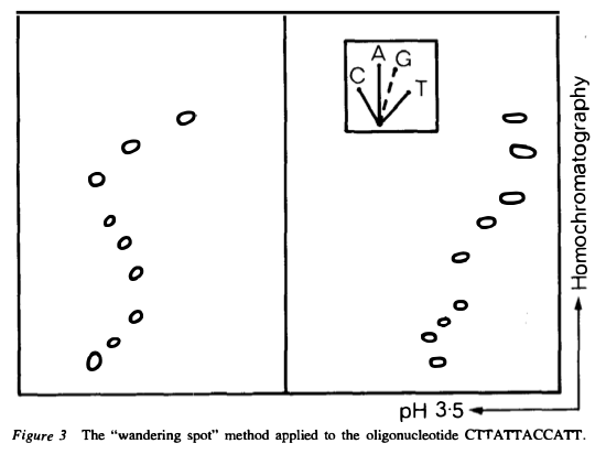
A powerful idea is partial hydrolysis of end-labeled material.

The sequence above can be read as TTACCCTT.  (This particular one is DNA).

lac operon control region

In 1975 the first natural DNA sequence was determined.


This was also the summer of the famous moratorium on recombinant DNA work.  It was obvious that a new era was beginning for biology.

Reznikoff et al. used a genetic trick in E. coli to transfer the lac operon's promoter/operator region onto lambda phage, transcribed across the control region in vitro, and then used a biochemical trick to enrich for sequences that were only from the lac control region.  The latter was to hybridize the RNA to separated strands of lambda phage without lac, and keep the stuff that didn't hybridize.

(PMID 1088926 --- this paper is locked behind a paywall at the journal Science, 45 years later, but I found a pdf linked on Bill Reznikoff's page at UW-Madison).

Maxam-Gilbert:  chemical method

In 1977 Wally Gilbert's lab introduced a chemical sequencing method for DNA.  I employed this method to sequence 70 bp of the promoter region for a late gene from phage T4 called gene 23.  I used to boast that mine was the shortest DNA sequence ever published but that's not actually true.  This one probably is.

The first step was to use alkaline phosphatase to remove 5' phosphates, then DNA kinase and 1 mCi of 32-P-ATP to label the ends.  If you do this with double-stranded DNA then both ends will be labeled.  You must somehow separate the strands or, cut with a restriction enzyme to get two different-sized pieces, and separate them by gel electrophoresis.  Here's a strand separation from the paper.


I used to do this on very thin acrylamide gels by denaturing the sample, loading it, and immediately cranking the power supply to 4000 V.  If you left it too long (> 15 sec), the heat would crack the glass plates.

The next steps were relatively simple organic chemistry
... the purines (A+G) are depurinated using formic acid, the guanines (and to some extent the adenines) are methylated by dimethyl sulfate [destabilizing the glycosidic bond], and the pyrimidines (C+T) are hydrolysed using hydrazine. The addition of salt (sodium chloride) to the hydrazine reaction inhibits the reaction of thymine for the C-only reaction. The modified DNAs may then be cleaved by hot piperidine; (CH2)5NH at the position of the modified base.
 What this does is to generate for each reaction a population of molecules, some of which end at the base in question.  So if the original molecule is

*NpNpApNpNpNpApNpNpNpApN

with * marking the 32-P, then after partial cleavage after A (adenosine) you have

*NpNpApNpNpNpApNpNpNpApN
*NpNpApNpNpNpApNpNpNpA
*NpNpApNpNpNpA
*NpNpA

Here is the wikipedia figure:


After gel electrophoresis and autoradiography (exposure of the gel to X-ray film), you get something like this:


And you can read the sequence.

Sanger sequencing

Fred Sanger developed a method for DNA sequencing.  It generates a population of molecules like above by using synthesis instead of degradation.  The synthesis is stopped short in some of the molecules by including a reagent which poisons the reaction, namely, a dideoxynucleotide triphosphate.

Here is the principle of the method in a figure from Sanger's (second) Nobel lecture.

In 1988, I spent about 3 months running about 100 sequencing gels to obtain about 3.5 kb of sequence data to determine the sequence of my favorite gene, hemA.  The data looked like this:


With somewhat better methods I spent about 6 months to obtain about 12 kb of sequence data in 1993-94.

Year  Amt         Time invested
1983  70 bp       months
1988  3500 bp     3 months
1994  12000 bp    6 months

Probably the biggest challenge in the method was to prepare the large polyacrylamide gels with no bubbles and a thickness at the top of 0.4 mm.

This era ended when commercial DNA sequencing services started.  Together with PCR, including PCR to amplify transposon-genome junctions, it revolutionized our work in the mid-1990s.  We would do a PCR reaction, send it in the mail, and 2 days later get a result by email.

Two very important adaptations were later made to the method.  First, rather than label the DNA, the dideoxy terminators were themselves labeled with fluorescent dyes.  Among other things, this meant that the reactions could be analyzed together in a single sample and analyzed by laser activation and a detector.

The other was replacement of polyacrylamide gels by capillary electrophoresis.  This extended the amount of information obtainable on one analysis of a sample from about 200 nt to about 1000 nt.

Machines do many reactions in parallel.  ABI 310.  You can buy a used one cheap, but caveat emptor.

That is the technology which was used to sequence the human genome, declared complete in 2003.


Reconstruction of SARS-CoV-2

A few days ago a paper was published in Nature after rapid review:

Rapid reconstruction of SARS-CoV-2 using a synthetic genomics platform.

It's quite amazing.  For starters, the work was completed in less than 6 weeks, since the first genomic sequence of the SARS-CoV-2 virus was released on Jan 10 and the paper was submitted on Feb 22.  Most of that time was preparation of the DNA fragments.

9 DNA fragments

The first step was to PCR amplify DNA segments spanning the genome.  This was done by RT-PCR.  Usually one verifies the sequence has not been mutated in the PCR reaction (I don't see this in the paper).  

Alternatively one can just order synthetic DNA of < 8 kb these days (with a 10 day turnaround)!  link NPR story

Cloning

Some DNA fragments are unstable when cloned into E. coli.  So they used YACs (yeast artificial chromosomes).  An additional advantage is that all 8 fragments can be assembled into the final product in one step!  (TAR-cloning)


In vitro transcription

The clones are made with a promoter for phage T7 RNA polymerase upstream and poly A tail downstream followed by a restriction site for Pac I.  YAC DNA was prepared, cut with Pac I and transcribed with T7 polymerase.

Transfection

The transcript was then transfected into mammalian cells, together with mRNA for one viral protein.  The system is a bit more complicated than that:  there are two cell types.  But it seems easy enough, and the result is that the transfected cell system produces virus.


The virus can then be cloned (classic terminology, progeny from a single individual produced) and grown in culture.  Simply amazing.

Attenuating mutations from viruses for the live-attenuated vaccine for SARS-CoV can be assembled into live SARS-CoV-2 and tested for growth properties and attenuation, and then investigated as vaccines.


Friday, May 8, 2020

Jonathan Swift

Falsehood flies, and the Truth comes limping after it.

Lewis Thomas on Streptococcal pneumonia

I've read a few case reports of COVID-19 cases. They talk about a slowly progressing disease, not particularly severe although the patients are miserable, until about day 10. At that time you get the severe pneumonia. I think it's probably a "cytokine storm", and I wanted to call it the "crisis", though I'm not sure that's an official term. Today I started to wonder about where I heard that before. And then I remembered:
"the patient complained of the sudden onset of chills and fever, cough, sometimes with blood-tinged sputum, and pain in one side of the chest; physical examination revealed dullness to percussion with one’s fingertips over the affected lung area and a characteristic change in the breath sounds heard with the stethoscope at the same spot. Given this amount of information you could begin making predictions. The prognosis for a young adult was the most surely predictable: an acute illness lasting ten to fourteen days, with a high fever each day, more chest pain and more cough, perhaps with alarming manifestations of exhaustion and debilitation near the end of this period, and then, suddenly and as triumphantly as the bright sunshine after a thunderstorm, one of the great phenomena of human disease — the crisis. On one day or another, after two weeks of his seeming to come closer and closer to death’s door, the patient’s temperature would drop precipitously within a few hours from 106 degrees to normal, and at the same time, with a good deal of sweating, the patient would announce that he felt better now and would like something to eat, and the illness would end, like that."
from Lewis Thomas, The Youngest Science.
Curious to know how they treated it? Streptococcal pneumonia was treated (1938) by first finding out what type of Streptococcus the patient had, and then injecting rabbit anti-Streptococcal serum. We wouldn't do that today, it's pretty risky. But I'm looking forward to convalescent patient serum, and perhaps humanized monoclonal antibodies for SARS-CoV-2.
If you've never read Lewis Thomas, you should. A biologist, an MD and a poet.

Originally from FB (2020-03-29).

Remdesivir

You've probably heard about remdesivir, it's all over the news.  Tony Fauci was on TV being excited about a not very exciting study the other day that "showed promise."

I've been quite interested in this drug ever since I read in NEJM that the first patient to be diagnosed as SARS-CoV-2 positive in the US ("Snohomish"), was treated under an EUA with remdesivir when they thought he was going south, and the next day, he got better.  Much better.  Supplemental oxygen was discontinued that day.  There is plenty of anecdotal evidence on the web in addition to Snohomish.  I read a piece about an ER doc the other day but I can't find it now.  The patient said "that magic juice works, Doc."

Remdesivir has a weird name.  Many drugs do. In this case, it's a result of a naming convention that antivirals end in -vir, monclonal antibodies in -mab and so on.  It's a very useful convention because one can immediately tell the class of a drug from its name.  There's a whole list of them here.  I don't know that remdes- is significant itself.

Like many antiviral drugs, remdesivir is a nucleoside analog.  Here it is compared with ATP (remdesivir on the left, adenosine triphosphate on the right).


Remdesivir was initially developed for Ebola and Marburg viruses (filoviruses).  It has broad antiviral activity (in vitro) for other virus families including paramyxoviruses and coronaviruses such as the first SARS-CoV.  These viruses are all RNA viruses, and remdesivir inhibits the enzyme that replicates the genome, which is an RNA polymerase.

As part of its clinical testing for Ebola, the safety profile in humans is known to be good, although there are some things to watch for.

As with all antivirals, it is likely to be most effective if given early in the disease.  The problem with that is that there doesn't seem to be much of it.  The maker, Gilead Sciences, just contributed its entire stockpile of 1.6 M doses to the federal government, and they are busy sending most of it states that voted for Trump (I'm not kidding).

Remdesivir is apparently hard to make.  Here is an article about some chemists who were able, with a lot of effort, to synthesize one gram of it.  Although you can take that with a grain of salt (or a shot of tequila).  This story has twists and turns in it.

Gilead

So Gilead is a company started in the late 1980s, the time when many people including my former professors John Abelson and Mel Simon had a startup to find drugs active against reverse transcriptase.  It is named after the Balm of Gilead.

Gilead developed a couple antivirals in the 1990s, and then (small world department) acquired a company called NeXstar Pharmaceuticals in Boulder (this is Larry Gold's company, another colleague from the phage T4 world).  If I'm reading wikipedia right, NeXstar was their key acquisition because they obtained a sales and marketing ability for Europe and other markets.

In any event, Gilead is the fish that keeps eating other fish its size or even a little larger, exactly the right ones, and then gaining weight.  Big enough to give away a lot of money.  According to the wikipedia article, "Charitable donations to HIV/AIDS and liver disease organizations totaled over 440 million in 2015."

One of the companies they acquired had developed sofosbuvir, an antiviral for Hepatitis C virus.  They paid 11 B and yet Forbes calls it ""one of the best pharma acquisitions ever".

You may have heard about these treatments.  HCV (hepatitis C virus) is essentially a death sentence.  Gilead's drug will cure you, but it'll cost you your house.

the cat story

So finally, there's a weird tie-in with cats, which I read this morning in the Atlantic.  (In fact, I've been so impressed with them, especially recently, that I purchased my first magazine subscription since Mad magazine).

Apparently remdesivir has a cousin, a closely related drug called GS-441524, also developed by Gilead.  It was discovered by a research scientist working on the disease FIP, that GS-441524 cures cats of this fatal GI illness, which is caused by a coronavirus.  Apparently, in spite of the pleas of legions of cat-lovers, Gilead will not try to license GS-441524 for this purpose, apparently because
any adverse effects uncovered in cats might have to be reported and investigated to guarantee remdesivir’s safety in humans.
So there is a shady black market in China for GS-441524.  I kid you not.  Read the story, it's great stuff.

Summary

This is just my two cents, which means little.  I am highly skeptical that a vaccine can be successfully developed in 18 months, let alone 6.  As I said in another post today, the optimistic experts say "maybe we will get lucky."

But I am very optimistic about therapeutics and particularly remdesivir, given early.  The thing about COVID-19 is that the lung damage is so severe.  Even if you survive it takes a long time to heal.  And antivirals always work best given early.  See oseltamivir (another Gilead drug).

DNA and RNA vaccines for SARS-CoV-2

People ask about the prospects for a vaccine for SARS-CoV-2.

The NYT had a good article about it about a month ago.

The history of vaccine development is a series of long drawn-out and frequently failed attempts.  The idea that we throw everything at this problem and it's just going to work, is pie-in-the-sky.  The optimistic experts say "maybe we will get lucky."

The proposals that would move the fastest are for DNA and RNA vaccines.  These are vaccines that would contain (for example) the gene for the Spike surface protein of the virus, with accessory elements giving high expression.

They would either be mRNA (modified to be resistant to breakdown) or DNA.

But you should be aware that no DNA or RNA vaccine has been licensed for humans, period.  There is one for West Nile Virus in horses.  Many have been tried but they frequently don't elicit neutralizing antibodies.

One problem is efficiency.  The nucleic acid just doesn't get into cells very well.  People are trying gene "guns" to improve efficiency:  the nucleic acid is bound to small metal microbeads, and propelled into your arm with compressed helium.

Moderna is an RNA vaccine that is in ongoing Phase 1 clinical trials, and has been approved to start Phase 2 even though the results from Phase 1 aren't known yet.  Phase 3 is expected to begin in early summer.

Summary

Phase 1:  Small N.   Is the vaccine grossly toxic or relatively safe?
Phase 2:  Medium N.  Is the vaccine effective?
Phase 3:  Large N    Is the vaccine very safe and also effective?

Analysts are even wondering where they are going to get all the vials they will need to put the vaccine in, and the syringes and needles for injections.  It's a real problem.


Thursday, May 7, 2020

Links to recent posts inspired by the pandemic

Covid-19 pathogenesis and epidemiology:
link Unsupported claim of increased pathogenicity (2020-04-23)
link Covid-19 deaths by county (2020-04-29)
link South Carolina cases (2020-04-30)
link Covid-19 CFR (2020-05-01)
link SARS-CoV-2 transmission (2020-05-03)
link Covid:  good news, bad news (2020-05-05)

SARS-CoV-2 virology:
link Phylodynamics, NextStrain, and "Snohomish" (2020-04-30)
link Rapid reconstruction of SARS-CoV-2 using a synthetic genomics platform

General virology:
link Introduction to animal viruses
link Measles virus

Vaccines:
link Overview of vaccination (2020-05-07)
link DNA and RNA vaccines for SARS-CoV-2 (2020-05-08)
link Virus attenuation (2020-05-18)

Therapeutics:
link Remdesivir

Biochemistry: 
link Covid tests:  PCR (2020-05-06)
link Covid tests:  LAMP + CRISPR (2020-05-06)
link Sensitivity, specificity and Bayes (2020-05-06)
link DNA sequencing:  classical methods (2020-05-08)
link Chromatography (2020-05-10)
link Electrophoresis (2020-05-10)

Literature:
link Lewis Thomas on Streptococcal pneumonia (orig FB 2020-03-29)

Stupid stuff debunked here (links):
link Lyme disease is a U.S. government-developed bioweapon (false)
link Mouse retrovirus associated with Chronic Fatigue Syndrome (false)
link Vaccines contain heavy metals (other than mercury) (false)
link Judy Mikovits (crazy person) + this
link Didier Raoult and HCQ (crazy person)
link No saline placebo trials for vaccines (false) --- No true Scotsman fallacy

A connection between thimerosal in vaccines and autism is refuted by systematic studies but also by the simple fact that since the removal of thimerosal from childhood vaccines, rates of autism diagnosis have not decreased as predicted but actually increased.

Overview of vaccination

A month ago there was an article in Nature that summarized current efforts toward a vaccine for SARS-CoV-2.  A total of 115 different projects were identified, 73 of them clearly moving forward.  Not much specific is publicly known as yet about these different projects.

In this post, I want to start with some history about vaccination and a broad overview of vaccine strategies.

We'll take a look at specific projects for Covid in another write-up.  I'm particularly interested to read up on modern approaches and what their chances of success are.

Variolation

Smallpox has been a greatly feared viral disease that was eliminated from the earth in the 1970s.  This guy is the last person to ever have smallpox.


(Except that the Russians kept samples, and apparently that lab had a fire recently).

Both Washington and Lincoln had smallpox, Washington as a young man when he visited the West Indies with his tuberculous brother Lawrence, and Lincoln about the time of the Gettysburg address.

Variolation involves taking pus from a smallpox patient and inoculating it to an uninfected person using a needle.  Often the resulting case would be mild.  It is an old practice dating back at least to the 1500s if not before.  It was brought to England in the early 18th century.
Maitland conducted an experimental variolation of six prisoners in Newgate Prison in London. In the experiment, six condemned prisoners were variolated and later exposed to smallpox with the promise of freedom if they survived. The experiment was a success, and soon variolation was drawing attention from the royal family, who helped promote the procedure throughout England.
The problem with variolation was that sometimes the smallpox that developed was severe.  This led Jenner to his famous development of the first vaccination (from the Latin vaccus, cow).  Supposedly he noticed that milkmaids had clear complexions, completely lacking the smallpox scars that were quite common for other people.  About 1798, he took samples of a virus related to smallpox, cowpox, and used that for inoculations with great success.


Vaccines

Classical vaccines are of three types.  The first to be developed were inactivated proteins.  These were made from protein (toxins) secreted by pathogenic bacteria as they are grown in culture.  [link]  The toxins are collected from the supernatant, concentrated and inactivated with formaldehyde.
Diphtheria once was a major cause of illness and death among children. The United States recorded 206,000 cases of diphtheria in 1921, resulting in 15,520 deaths. Diphtheria death rates range from about 20% for those under age five and over age 40, to 5-10% for those aged 5-40 years. Death rates were likely higher before the 20th century. Diphtheria was the third leading cause of death in children in England and Wales in the 1930s. 
Since the introduction of effective immunization, starting in the 1920s, diphtheria rates have dropped dramatically in the United States and other countries that vaccinate widely. Between 2004 and 2008, no cases of diphtheria were recorded in the United States. 
There is a famous dog named Balto, part of a sled team that brought the diphtheria vaccine to Nome, Alaska during an outbreak in the 1920s.  I'm not sure he'd be happy about it but Balto was stuffed and currently resides in a museum in Cleveland.


[Update:  the treatment for diphtheria was not a vaccine, but an "antitoxin", that is, antibodies to the toxin protein.  Antitoxins are commonly made in large animals, especially horses.  There is a vaccine, but that came later. ]

The other types of vaccine use the actual infectious agent.  There is a vaccine for tuberculosis that is an attenuated strain of Mycobacterium tuberculosis called BCG.

A vaccine against a virus like polio may be inactivated or alternatively, a live but attenuated virus.  These vaccines depended originally on the development of mammalian cell culture and its use to produce virus in quantities large enough to immunize large numbers of people.

Inactivation is by treatment with chemicals (e.g. formaldehyde) or possibly UV.  Attenuation means that the virus grows, but does not cause disease, either because it grows too slowly, or because it is unable to infect a particular tissue (poliovirus in the nervous system).  Finding an attenuating mutant is a laborious process.  In testing pathogenicity, there is no substitute for tests with live animals.

Besides tetanus, the first group of toxoid vaccines includes diphtheria.  An example of the second is the Salk polio vaccine, and the third class includes many important vaccines including

- Yellow fever
- MMR (measles, mumps and rubella)
- Varicella (chickenpox)
- the oral (live) Sabin polio vaccine
- live (nasal spray) influenza vaccine
- Smallpox

In the last 30 years or so, vaccines have been developed for Streptococcus pneumoniae using its capsular polysaccharide, which may be chemically attached (conjugated) to a protein to make it more immunogenic.  The capsule is important in preventing engulfment (phagocytosis) by cells of the immune system called neutrophils.  Antibodies to the polysaccharide capsule allow phagocytosis and prevent disease.

Most viruses are first encountered by the host on mucosal surfaces in the mouth, the gut (digestive tract) or the airways (respiratory tract).  In these environments a particular type of antibody called IgA is predominant.

Vaccine dogma holds that to produce strong immunity, a good IgA response is essential.  The vaccine type which is best at achieving a good IgA response is thought to be an attenuated live virus vaccine, because it is the gift that keeps on giving.  Continuous presence of the antigen drives antibody producing cells (B-cells) to switch to making IgA.

Three vaccines

IPV

The news of the success of the killed polio vaccine developed by Jonas Salk was greeted with joy in 1955.  This vaccine is called the Salk vaccine or IPV (inactivated poliovirus vaccine), and it's based on three wild, virulent reference strains:  Mahoney (type 1 poliovirus), MEF-1 (type 2 poliovirus), and Saukett (type 3 poliovirus).  The three viruses are grown in tissue culture (Vero cells, a type of monkey kidney cell), and then collected and inactivated with formaldehyde (formalin).

Salk's approach was widely criticized as dangerous, and indeed, in that same year a batch from the Cutter Laboratories in Berkeley, California was not properly inactivated and it resulted in 200 cases of polio and 11 deaths.

OPV

A different vaccine using attenuated live virus was developed by Albert Sabin, called the oral polio vaccine (OPV).  It came into use around 1961.  The OPV contains the three prevalent serotypes of poliovirus which have been passaged for many generations in tissue culture and accumulated mutations that do not interfere too much with growth in the gut, but prevent neurovirulence, growth in the nervous system.  The individual components of the MMR vaccine were also developed in the 1960s.

It seems that the main reason the oral polio vaccine won out at that time was that the Salk vaccine is an injection, while the live virus is administered orally, often a drop or two on a sugar cube.  Kids prefer the sugar cube.  Also, sterile syringes are in short supply around the world.  As an attenuated live virus, the Sabin vaccine is expected to be better at inducing a good IgA response.  There is also evidence that the Sabin vaccine interferes with shedding of live poliovirus when they are both present, causing the live virus to die out.


The attenuated virus in the Sabin vaccine has been sequenced (years later).  Although there had been speculation about deletion mutations, apparently there are just a number of single nucleotide substitutions.  Only a few of these are responsible for the attenuation phenotype in any given vaccine strain.  [review]

It happens rarely that the OPV causes authentic polio, a phenomenon called VAPP (vaccine-associated paralytic polio).  The sequence of some VAPP strains shows reversion of the mutations in a one of the vaccine strains to change at least some of the attenuating mutations back to wild type.  Also, it turns out that there are other naturally occurring viruses in the same family as poliovirus (Picornaviridae, for small RNA virus), which occur in nature and these may sometimes recombine with the live Sabin virus to give a recombinant that has recovered the ability to grow well in the nervous system (ref).

In any event, this reversion is problematic not so much for the vaccine recipient (who is well on the way to immunity) but for other residents of an area with poor sanitation.  The transmission of vaccine strains to others in poor communities was originally held to be a feature, not a bug.

VAPP, which is extremely rare, finally became of crucial importance when polio was eliminated from most parts of the world.  Then, having a risk of developing authentic polio out-weighed the advantage of the oral vaccine.  When our son was born, we wrestled with the question of which vaccine to use for immunization.  We chose Salk's IPV.  Later, the vaccine schedule was changed to have the killed virus first and the live virus for boosters.

Here is a slide that shows the difference between IPV and OPV in the IgA response.


Although an IgA response is desirable, it is obviously not essential.  IPV works well.  That may well have something to do with the real pathogenesis happening in neural tissue.

Flu

The third vaccine to talk about is the live influenza vaccine.  As a virus, influenza is unusual because it has a segmented genome, each gene is on a separate piece of RNA within the virus particle.

In epidemiology, influenza is different than most viruses because it mutates rapidly enough to escape the immune response within two or three years.

As with all viruses, a major component of the immune response is directed against proteins (antigens) on the surface of the virus.  For influenza these are HA (hemagglutinin) and NA (neuraminidase).


HA (hemagglutinin) is a viral protein that binds to the receptors for influenza virus on animal cells.  These receptors are proteins with sugar chains attached to them and with the special sugar sialic acid attached at the end.  HA binds to sialic acid.  There are subtle differences in the structure of different forms of sialic acid between tissues or say, comparing human and avian hosts.  Some HA bind better to human cells.  HA is sometimes further abbreviated as H.

There is a second surface protein called NA (neuraminidase), further abbreviated N.  Its role is to cleave sialic acid from the sugar chain when newly synthesized virus is leaving the cell.  Different strains of influenza virus have different types of H and N.

The influenza virus from the 1918-20 pandemic, that has circulated in various permutations ever since, is H1N1.  The virus of the 1956-57 pandemic was H2N2, and that for 1968-69 was H2N3.

This antigenic drift means that a new vaccine must be formulated to best match the viruses that are circulating in any particular year.  The segmented genome makes it fairly easy to produce a new vaccine strain by coinfecting cells with the virus whose HA and NA you want, plus the old vaccine strain.  Reassortment of viral genome segments produces different combinations which can be screened to find the desired type that has attenuation mutations but the target HA and NA.


However, this also means that it is fairly easy (in evolutionary time) for completely new HA and NA genes to be transferred from avian flu strains to human flu strains.  This is called antigenic shift, and it happened successfully in 1918, 1956 and 1967.  The 2009 virus came from pigs (although it must be noted that we gave it to them first, in 1918, so it's not really fair to complain).

Some more slides from one of my old lectures:


Other approaches

HPV (Human papilloma virus) comes in many serotypes.  These viruses cause warts, but certain types are also associated with various cancers.

HPV is a small DNA virus and the proteins of the virus can self-assemble into a virus particle.  One HPV vaccine is made from hollow "virus-like-particles" assembled from proteins made by recombinant DNA methods, without the genome inside.

There are other approaches to making vaccines that have the promise of more rapid scale-up including DNA and RNA vaccines, and recombinant proteins (e.g. the S or Spike protein).  Also of importance are viruses that have been modified to not be harmful but can be used as carriers to express the desired cornavirus protein antigens.  These include adenovirus and retrovirus-based vaccines.

For many of these, there is a concern that they will not boost the immune system as much as is necessary to give a good response.  Various compounds (or even killed bacterial cells) act as adjuvants, treatments that increase the immune response.  Many different combinations are possible and probably most of them are being tried.

It is noteworthy that the subunit vaccine (recombinant proteins) for pertussis (whooping cough) doesn't seem to be as active as the old vaccine, which had the drawback that most kids had soreness and redness at the site of the injection for several days.

Failures

We do not yet have successful vaccines for several important targets.  These include HIV and malaria.  In the case of HIV, the success of therapeutics may have something to do with the lack of progress on a vaccine, but it is a problem where many approaches have failed.

Risks

There are a couple of negative points to mention about vaccines.

One is that it is important to keep the vaccine formulation sterile.  A tragic early event was the death of some Australian children due to injection with diphtheria vaccine that was contaminated with Straphylococcus aureus (the Bundaberg disaster).  The famous Australian scientist Macfarlane Burnet had a role in solving this mystery.

A second issue is that occasionally, for a particular vaccine, the immune response makes an infection with the virus worse.  The example we have of this is a Dengue called Dengvaxia.  However, this hasn't been seen with other viruses, or even other vaccines for Dengue.

Thimerosal

Finally, there is the issue of Thimerosal.  After problems with vaccine preparations which had been stored and allowed bacterial growth, it became the practice to add low concentrations of a compound containing mercury called thimerosal.

In the 1990s anti-vaccine activists popularized the notion that thimerosal in vaccines may have a connection to autism.  In response, thimerosal was removed from vaccines for children by 2001.  Rates of autism haven't changed, and there was never any evidence connecting thimerosal to autism.  One prominent activist, Andrew Wakefield, was found guilty of scientific fraud.  (He was motivated by payments by a group of lawyers who hoped to profit by suing vaccine manufacturers).

Non-human vaccines

There are a number of important vaccines for animals, including pets.  The "core" vaccines recommended for all dogs are rabies, distemper, parvo, and adenovirus.


Wednesday, May 6, 2020

Sensitivity, specificity and Bayes

Now that we're ramping up testing, including serology, you're going to see words like sensitivity and specificity thrown around. Here's a brief primer.

Rather than give a bunch of definitions right away, I would rather start by asking a question and then showing how we can answer it by building a table, based on three numbers (really, ratios).

I just got a positive test back for X.  Do I really have X? 

We have to recognize that there is some uncertainty here.  Peter Norvig:
suppose I have a test machine that determines whether the subject is a flying leprechaun from Mars. I'm told the test is 99% accurate. I put person after person into the machine and the result is always negative (correctly). Finally one day, I put someone (say, Tom Hanks) into the machine and the light comes on that says "Flying Leprechaun!" Would you believe the machine? Of course not: that would be ridiculous, so we conclude that we just happened to hit the 1% where the test errs. 
We find it easy to completely reject a test result when it predicts something impossible (even if the test is very accurate); now we have to train ourselves to almost completely reject a test result when it predicts something almost completely impossible (even if the test is very accurate).
I used the following example for my students:  a person walks into the doctor's office with a very bad sore throat and a rapid Strep test (for Streptococcus pyogenes) is administered.  A positive result is obtained.  What is the probability that the patient really has a strep throat?

Let's construct a table:
                        strep    No strep   total
positive test                                      pos tests
negative test                                      neg tests
total w/ or w/o strep

The question we are asking is equivalent to the following:  take the number of people with a positive test that have Strep (that's the first number in row 1) and divide by the total number of people with a positive test (the third number in row 1).

Well, how do you do that?

Sensitivity is an umpire who sees a real strike and calls "strike."  It is the proportion of people who actually have the disease that also have a positive test result.  For rapid Strep tests, this number is about 90-95%, let's call it 90.  So out of every 100 people, we write

                        strep    No strep   total
positive test              90                       pos tests
negative test              10                       neg tests
total w/ or w/o strep     100                           

Another (compound) word to describe those 10 people who have Strep but got a negative test is false-negatives.

Specificity is an umpire who sees a pitch that is actually a ball, and calls "ball".  It is the proportion of people who do not have the disease and have a negative test result.  For rapid Strep tests, this number is claimed to be about 98%.
                        strep    No strep   total
positive test              90          2            pos tests
negative test              10         98            neg tests
total w/ or w/o strep     100        100                 

Those 2 people who have Strep but got a negative test are false-positives.

Now, to get the total number of positive tests can I just add the two values in the first row?  No!!!

The reason is that the totals on the bottom need to be adjusted.  In our table so far, we have equal numbers of people that have Strep or don't have it.  

But in the real world, the actual proportion of people who walk in complaining of sore throat that have Strep is about 10%.  This is called the prevalence (or incidence if you want to be picky).  It is determined by doing further tests (streaking a throat swab onto blood agar) and other things such as PCR.

To fix this, we scale all the values in column 2 so that the total on the bottom is 900.  Then the prevalence will be right.  We do that by multiplying each value by 9.

                        strep    No strep   total
positive test              90          18     108   pos tests
negative test              10         882     892   neg tests
total w/ or w/o strep     100         900    1000         

So the probability that you actually have Strep, given a positive test, is about 90/108 = 83%.

Now, you may think this is no big deal.  83% is still a lot, even if it's not 90 or 98%.  But this is a very good test, and the prevalence is reasonably high.

Suppose we have a different example with the same sensitivity and specificity but the prevalence is 1%.

                      disease  No disease   total
positive test               9          20      29   pos tests
negative test               1         970     971   neg tests
total w/ or w/o disease    10         990    1000         

That's not so good.  Now only one-third of people who receive a positive test result actually have the disease, even with a very good test.

Sensitivity is about the ump calling strikes correctly. Specificity is about the ump calling balls correctly. Prevalence is how often the pitcher throws a strike.

Prevalence has a big influence and this is often forgotten. Since the prevalence of people who have had COVID is extremely low (first written early April, 2020), even sensitivities and specificities in the high 90s will be problematic for population-level serology testing. I leave making the table for that as an exercise...

What's the connection to Bayes?  Bayes theorem leads to a system for probability which we can think of as starting with some prior likelihood for a particular statement and then updating as evidence becomes available.

In this case our prior for the hypothesis that a patient has the disease is the prevalence.  When updated by the test result we get the final probability.

[ To do the math more easily, start with the population and first calculate disease/no disease using the prevalence.  Then use the sensitivity and specificity to get the other numbers. ]